AQA GCSE Using Ultrasound Waves for medicine(Physics)
Ultrasound Waves
Ultrasound waves are very high frequency sound waves.
Frequency of ultrasound waves is >20,000Hz, so humans cannot hear ultrasound waves.
We have many different uses for ultrasound waves, they are used for pre-natal scans, or for scanning body organs.
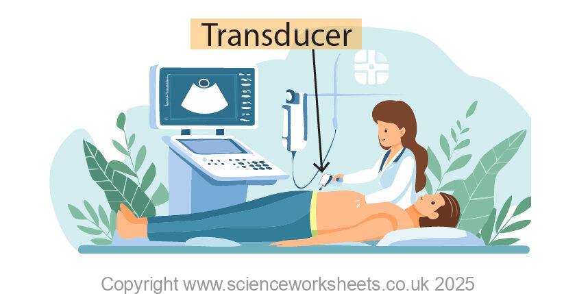
In the image above the hospital worker is using the transducer of the ultrasound to scan the patients body. The transducer will emit and detect pulses of ultrasound waves as shown below.
The pregnant mother has removed her jumper from her stomach to allow the transducer to be placed onto the skin of her stomach.
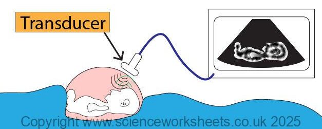
Transducer will emit ultrasound waves (black colour) these pass through tissue layers. At each boundary some of the ultrasound wave is reflected back (green wave).
The transducer will receive these reflected waves, they are processed to form an image of the baby on the screen.
The computer that forms part of the ultrasound will record the time for the reflected sound wave to reach the transducer and using speed of sound it will calculate the distance of the structures in the scanned image.
The image formed on the screen is a 2d image, but the hospital operator will move the transducer across the area in a sweeping motion so they can see from different view points and build up an overall image.
Ultrasound or X-rays?
Both ultrasound and X-rays can be used for imaging in hospitals. There are reasons for choosing a particular method.
| Ultrasound | X-rays | |
|---|---|---|
| Produced by | High frequency vibration | X-ray tube |
| Ionising | Non-ionising | Ionising |
| Potentially harmful | Non harmful | Slight risk of harm as they are ionising, but benefits often outweigh risks |
| Used for | Provides images of soft tissues | Bone fractures. When used for soft tissues a contrast medium such a barium meal needs to be taken. |
Practice Question
1.State what ultrasound waves are
2.What would be the expected frequency of an ultrasound wave?
3. State two possible medical uses for ultrasound waves.
4. State the name of the piece of apparatus that the hospital worker uses during an ultrasound scan which emits and detects ultrasound waves.
5. Explain how an ultrasound machine is able to produce an image which shows soft tissues.
6. Suggest why many doctors will use ultrasound as an imaging method over x-rays.
Absorption and Emission of EM Radiation
JJ Thomson and Plum pudding model
Ernest Rutherford and the Nuclear Model
Niels Bohr changing the Nuclear Model
Discovering the Proton and Neutron
Measuring radiation from radioactivity
Radiation types and properties
Random nature of radioactive decay
Radioactive contamination or irradiation
Hazards of contamination and irradiation
Studies on the effects of radiation on humans
Different half lives of radioactive isotopes
Nuclear Fission Chain Reaction
Writing nuclear fission equations
Drawing ray diagrams for a concave lens
Drawing Ray Diagram to produce a virtual image for a convex lens
Drawing ray diagram to produce a real image for a convex lens.
Specular and Diffuse Reflection
Seeing Coloured Objects Part 2
Viewing objects through coloured filters
Transparent, Translucent and Opaque