Answers to AQA GCSE Microscopes(Biology)
Practice Question
1.Define the term magnification
Magnification = how much bigger the image is compared to the object.
2.Define the term resolution
Resolution = how clearly two points can be distinguished as separate.
3. Explain how the development of electron microscopes have allowed us to advance our understanding of cell structure.
Light microscopes (earlier development)
Invented first and let scientists see cells for the very first time (Hooke, Leeuwenhoek).
But their maximum magnification is only around ×1,500–×2,000.
Their resolution (the ability to distinguish two points as separate) is limited to about 200 nm.
This meant only larger structures could be seen — nucleus, cell wall, cell membrane, cytoplasm, chloroplasts.
Electron microscopes (20th century)
Two main types: Transmission Electron Microscope (TEM) and Scanning Electron Microscope (SEM).
Use beams of electrons instead of light, which have a much shorter wavelength.
Magnification: up to ×2 million or more.
Resolution: around 0.1 nm (much higher than light microscopes).
How this advanced understanding of cells
Allowed scientists to see much smaller sub-cellular structures in detail, such as:
Ribosomes (where proteins are made)
Mitochondria (internal folds/cristae for respiration)
Internal structure of chloroplasts (grana, thylakoids)
Fine details of cell membranes
Enabled us to better understand how cells function, e.g. energy production, protein synthesis, transport across membranes.
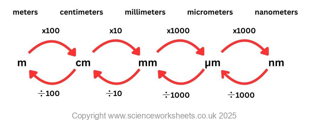
4. A bacterial cell has a diameter of 1μm. When drawn on a piece of paper it is 4cm. Calculate the magnification of the image.
Magnification = image size / size of real object
Actual size: 1 μm = 0.0001 cm
Image size: 4 cm
Magnification= 4 cm / 0.0001 cm = 40,000
Answer: = (×40,000).
Factors affecting the rate of photosynthesis
Measuring & calculating rates of photosynthesis
Inverse square law and photosynthesis
Economics of enhancing the conditions in greenhouses
Investigating the effect of light intensity on the rate of photosynthesis
Data analysis and nervous system
The effect of a factor on human reaction time
Controlling blood glucose concentration
Regulating water and nitrogen levels in the body.
Hormones and human reproduction
Using hormones to treat infertility
Control and coordination in plants using hormones
Investigating the effect of light or gravity on the growth of newly germinated seedlings